Team UICMA
Responsible for UICMA: DRA. Ana Riveros Salvatierra
Academic team: Dr. Felipe Oyarzun – Dr. Marcelo Kogan
This new service platform provides the possibility of characterizing comprehensively the various advanced materials, including nano and microstructures of organic and inorganic origin. It therefore encompasses all areas of basic and applied science. This service also is rigorously monitored by scientists who develop the characterizations, They advise on the interpretation of the data and report possible projections of the results
Its benefits are available to the scientific community and public and private research centres.
Our platform has team of high-tech for the characterization of: morphology, topography, size, elemental composition, hydrodynamic diameter, potential z and quantification of nano and microstructures organic and inorganic
Equipment available:
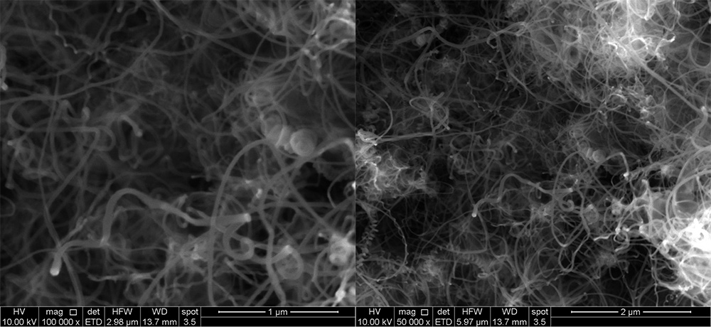
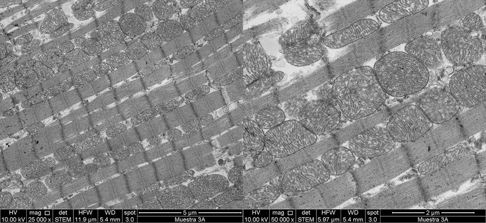
FE-SEM, High resolution scanning electron microscope (High Resolution Scanning Electron Microscope): Allows generating high-resolution images (1-3 nm), due to its focused electron beam by field emission. This team has with:
- STEM, Transmitted electron detector (Scanning Transmission Electron Microscopy, In English): It detects the electrons transmitted through the sample, to build a 2D image, delivering information about the shape and size of the particle. With the additional advantage of reducing damage by radiation, Since the beam moves sweeping the sample. It allows you to see cuts of tissues and cells, for studies in cell biology, Molecular and biopsies for clinical analysis.
- IS, Secondary electron detector: Of low-energy secondary electrons (<50 eV) emitted from the surface of the sample can be used to generate a topographic image, Noting the relief of the sample study.
- Beam Deceleration, which allows to analyze samples at low voltage (4kV) to improve aberrations. This represents an advantage when analyzing biological samples, since improving its resolution and contrast without jeopardizing the integrity of the organic sample.
- DBS, Directional Back-scattered ultrasensitive detector. This detector features four concentric ring segments that allow the detection of electrons emitted at different angles. The four segments can be purchased at the same time, in this way it is possible to select multiple contrasts and improve the resolution of the sample.
- vCD: High-contrast sensor. Detector of high sensitivity solid state retrodispersado. Can be used with the Beam Deceleration, which allows to analyze samples at low voltage (4kV) to improve aberrations. This represents an advantage when analyzing biological and non-conductive samples, since improving its resolution and contrast without jeopardizing the integrity of the sample.
- Elementary chemical analysis by spectroscopy detector (Energy Dispersive Spectroscopy, EDS): This detector performs the elemental chemical analysis by x-ray energy dispersive spectroscopy x in high resolution images. It allows you to observe cells or tissue sections by contrast and identify the composition and spatial distribution of nanoparticles, In addition to the information that gives us, respect the integrity of the biological model used either tissue or subcellular level. Also, It allows to make a mapping of elemental composition of different surfaces nanostructured materials. This tool allows you to identify the elements present in the sample and determine the proportion of each element and its spatial distribution. These analyses can be made both with the detector is and STEM.
Zeta Sizer nanoZS: Allows the determination of the molecular size of suspended particles from a nanometer to Micron, the dynamic scattering of light by setting the hydrodynamic diameter and zeta potential. The measurement can be performed in the native environment of the material.
NanoSight, Nano tracking analysis (NTA): NTA technology determines the concentration of nanoparticles and establishes a population distribution by composition. In addition, Displays the movement of Nanosystems in suspension its reactivity in static and/or subject to flow media, allowing to analyze, the capacity of the nanostructures to interact with other species in suspension or fixed (other particles, protein and substrate of interest) and dynamic training systems in response to various stimuli. The team also allows you to analyze the behavior of fluorescent structures of special usefulness in biotechnological applications.
Due to the great interest of the University of Chile for research, development and innovation (R & d), and the transversality of the study of nano and microstructures in disciplines basic and applied in the field of nanobiotechnology, nanomedicine and nanochemistry, We plan us UICMA is a national benchmark of nanoscience and a unit of collaboration and support in the development of new technologies.
MISSION AND VISION
Investigation and characterization of advanced materials unit aims to promote the scientific development of the University of chile and other public and private institutions, putting at the service of the equipment of high technology and specialized advising researchers.
In addition, provide support in the training of professionals and researchers from undergraduate and graduate.
At last, UICMA is projected to be a national reference both to the academic area, as the industry area, in order to encourage the development of new technologies at the national level.
AWARDED PROJECTS
FONDEQUIP EQM160157 "implementation of a platform for characterizing nanostructures (size, zeta potential, concentration and fluorescence) interest in different disciplines"
FONDEQUIP EQM170111 "strengthening of the area of electron microscopy for topographical characterization, size and elemental analysis of nanomaterials that are focused on the development of nanobiotechnology"
Contact
DRA. Ana Riveros Salvatierra
Phones: 229782918
E-mail: servicios.hrsem@ciq.uchile.cl
Direction: Santos Dumont 964, building Luis Nuñez, floor - 1
Author: Valentina Parra
Laboratory of molecular signal transduction of the ACCDiS and cell differentiation and metabolism laboratory; Facultad de Ciencias Químicas y Farmacéuticas; University of Chile.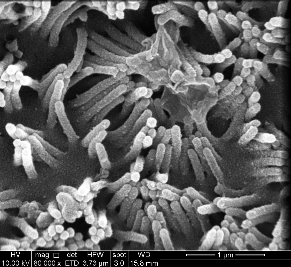
Author: Miltha Hidalgo, Ana Riveros, Porras Omar
Research laboratory in functional nutrition (LINF) of the University of Chile-INTA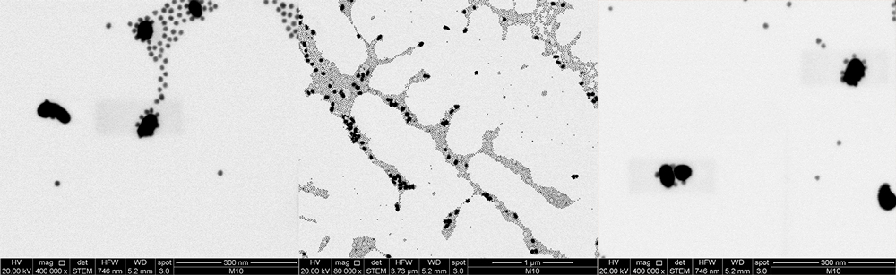
Authors: Maria Jose Marchant Lillo